Comprehensive Guide to Hip Anatomy and Conditions
The hip joint is where the upper body connects to the lower body. As such, it's critically important for walking, bending, and lifting, among others. All of the anatomical parts of the hip must work together to enable various hip movements. If any part of the hip has injuries, problems, or conditions, it can not only cause tremendous pain, but severely limit a person's ability to move and function. We're here to help your hip health, so you can have the foundation for a happy, pain-free life.
To help you discuss hip problems, conditions and treatment options with your surgeon, here's a breakdown of the anatomy of the hip.
Guía integral de la anatomía y las afecciones de la cadera
La articulación de la cadera es donde la parte superior del cuerpo se conecta con la parte inferior del cuerpo. Por lo tanto, es críticamente importante para caminar, doblarse y levantar objetos, entre otras funciones. Todas las partes anatómicas de la cadera deben trabajar en conjunto para permitir diversos movimientos.
Si alguna parte de la cadera presenta lesiones, problemas o afecciones, no solo puede causar dolor intenso, sino también limitar gravemente la capacidad de movimiento y funcionamiento de la persona.
Estamos aquí para ayudar con la salud de tu cadera, para que puedas tener la base para una vida feliz y sin dolor.
Para ayudarte a hablar sobre problemas de cadera, afecciones y opciones de tratamiento con tu cirujano, aquí tienes un resumen de la anatomía de la cadera.
- AnatomyAnatomía
- Conditions Condiciones
- Procedures Cirugías
Hip Anatomy
The hip joint is the largest weight-bearing joint in the human body, and is a ball and socket joint that is surrounded by muscles, ligaments, and tendons.
Bones of the Hip
The hip joint is the junction where the hip joins the leg to the trunk of the body. It is composed of two bones:
- The thigh bone, or femur
- The pelvis, which is made up of three bones called ilium, ischium, and pubis The hip bone is covered by a thin, tough, flexible and slippery surface called articular cartilage, and enables smooth movements of the bones and reduces friction.
Joints of the Hip
The ball of the hip joint is made by the femoral head, while the socket is made by the acetabulum. The acetabulum,muscles, and ligaments surround and support the hip joint.
Ligaments of the Hip
Ligaments of the hip joint provide stability to the hip by forming a dense and fibrous structure around the joint capsule. There are four ligaments adjoining the hip joint:
- Iliofemoral ligament is a Y-shaped ligament that connects the pelvis to the femoral head at the front of the joint to help in limiting over-extension.
- Pubofemoral ligament is a triangular shaped ligament that extends between the upper portion of the pubis and the iliofemoral ligament, and attaches the pubis to the femoral head.
- Ischiofemoral ligament is a group of strong fibers from the ischium behind the acetabulum that merge with the fibers of the joint capsule.
- Ligamentum teres is a small ligament that extends from the tip of the femoral head to the acetabulum, and has a small artery that supplies blood to the femoral head.
- Acetabular labrum is a fibrous cartilage ring that lines the acetabular socket, deepens the cavity, and increases the stability and strength of the hip joint.
Muscles and Tendons of the Hip Joint
The iliotibial band is a long tendon that runs from the hip to the knee, and is an attachment site for several hip muscles:
- Gluteals are three muscles (gluteus minimus, gluteus maximus, and gluteus medius) that attach to the back of the pelvis to form the buttocks.
- Adductors are located in the thigh which helps pull the leg back towards the midline.
- Iliopsoas is located in front of the hip joint and provides flexion.
- Rectus femoris is the largest band of muscles located in front of the thigh, and provides hip flexion.
- Hamstring muscles begin at the bottom of the pelvis and run down the back of the thigh, and help in extension of the hip by pulling it backward.
Nerves of the Hip
The main nerves in the hip region are the femoral nerve in the front of the femur and the sciatic nerve at the back. The hip is also supplied by a smaller nerve known as the obturator nerve.
Arteries of the Hip
The femoral artery, one of the largest arteries in the body, travels through the hip to supply blood to the lower limbs.
Suffering from hip injuries or conditions? Ready to feel what it's like to live free of hip problems and pain? Let us help your hips.
Anatomía de la cadera
La articulación de la cadera es la articulación de soporte de peso más grande del cuerpo humano, y es una articulación esférica (ball and socket) que está rodeada por músculos, ligamentos y tendones.
Huesos de la cadera
La articulación de la cadera es la unión donde la cadera conecta la pierna con el tronco del cuerpo. Está compuesta por dos huesos principales:
- El fémur, o hueso del muslo
- La pelvis, que está formada por tres huesos llamados ilion, isquion y pubis
- El hueso de la cadera está cubierto por una superficie delgada, resistente, flexible y resbaladiza llamada cartílago articular, que permite movimientos suaves de los huesos y reduce la fricción.
Articulaciones de la cadera
La cabeza femoral forma la esfera de la articulación de la cadera, mientras que el acetábulo forma la cavidad. El acetábulo, los músculos y los ligamentos rodean y sostienen la articulación.
Ligamentos de la cadera
Los ligamentos de la articulación de la cadera proporcionan estabilidad al formar una estructura densa y fibrosaalrededor de la cápsula articular. Hay cuatro ligamentos principales que se unen a la articulación de la cadera:
- Ligamento iliofemoral: es un ligamento en forma de Y que conecta la pelvis con la cabeza femoral en la parte frontal de la articulación para limitar la sobreextensión.
- Ligamento pubofemoral: es un ligamento triangular que se extiende entre la parte superior del pubis y el ligamento iliofemoral, y une el pubis con la cabeza femoral.
- Ligamento isquiofemoral: es un grupo de fibras fuertes que parte del isquion detrás del acetábulo y se fusiona con las fibras de la cápsula articular.
- Ligamentum teres: es un pequeño ligamento que se extiende desde la punta de la cabeza femoral hasta el acetábulo, y contiene una pequeña arteria que suministra sangre a la cabeza femoral.
- Labrum acetabular: es un anillo de cartílago fibroso que recubre la cavidad acetabular, profundiza la cavidad y aumenta la estabilidad y fuerza de la articulación.
Músculos y tendones de la articulación de la cadera
La banda iliotibial es un tendón largo que va desde la cadera hasta la rodilla y sirve como sitio de inserción para varios músculos de la cadera:
- Glúteos: son tres músculos (glúteo menor, glúteo mayor y glúteo medio) que se unen en la parte posterior de la pelvis para formar los glúteos.
- Aductores: están ubicados en el muslo y ayudan a llevar la pierna hacia la línea media del cuerpo.
- Iliopsoas: está ubicado en la parte frontal de la articulación de la cadera y proporciona flexión.
- Recto femoral: es la banda muscular más grande ubicada en la parte frontal del muslo y proporciona flexión de la cadera.
- Músculos isquiotibiales: comienzan en la parte inferior de la pelvis y descienden por la parte posterior del muslo, ayudando en la extensión de la cadera al tirarla hacia atrás.
Nervios de la cadera
Los nervios principales en la región de la cadera son:
- Nervio femoral: ubicado en la parte frontal del fémur
- Nervio ciático: ubicado en la parte posterior
Además, la cadera está inervada por un nervio más pequeño conocido como nervio obturador.
Arterias de la cadera
La arteria femoral, una de las arterias más grandes del cuerpo, atraviesa la cadera para suministrar sangre a las extremidades inferiores.
¿Sufres lesiones o afecciones en la cadera?
¿Listo para sentir lo que es vivir libre de problemas y dolor en la cadera?
Déjanos ayudarte con tus caderas.
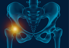
Hip Pain
Hip pain, one of the common complaints, may not always be felt precisely over the hip joint rather in and around the hip joint. The cause for pain is multifactorial and the exact position of your hip pain suggests the probable cause or underlying condition causing it.
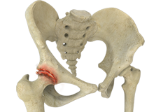
Hip Arthritis
Osteoarthritis, also called degenerative joint disease, is the most common form of arthritis. It occurs most often in the elderly. This disease affects the tissue covering the ends of bones in a joint called cartilage. In osteoarthritis, the cartilage becomes damaged and worn out, causing pain, swelling, stiffness and restricted movement in the affected joint.
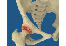
Rheumatoid Arthritis
Rheumatoid arthritis is a chronic inflammatory disease in which the lining of your joints becomes inflamed, causing pain, swelling, and stiffness. It is an autoimmune disease because it occurs when your immune system, which normally fights against infection, starts destroying healthy joints.

Dolor de cadera
El dolor de cadera, una de las quejas comunes, no siempre se siente exactamente sobre la articulación de la cadera, sino en y alrededor de la misma. La causa del dolor es multifactorial y la posición exacta de su dolor de cadera sugiere la causa probable o la condición subyacente que lo provoca.

Artritis de cadera
La osteoartritis, o enfermedad articular degenerativa, es el tipo más común de artritis. En la osteoartritis de la cadera, el cartílago que recubre los extremos de los huesos en la articulación de la cadera se daña y desgasta, causando dolor, hinchazón, rigidez y movimiento restringido.

Artritis reumatoide
La artritis reumatoide es una enfermedad inflamatoria crónica en la que el revestimiento de sus articulaciones se inflama, causando dolor, hinchazón y rigidez. Es una enfermedad autoinmune porque ocurre cuando su sistema inmunológico, que normalmente lucha contra las infecciones, comienza a destruir articulaciones sanas.
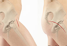
Minimally Invasive Hip Replacement
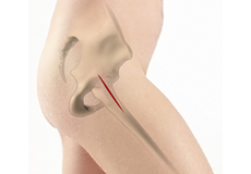
Minimally invasive total hip replacement is a surgical procedure performed through one or two small incisions rather than the single long incision of 10-12-inches as in the traditional approach.
Anterior Hip Replacement
Direct anterior hip replacement is a minimally invasive hip surgery to replace the hip joint without cutting through any muscles or tendons as against traditional hip replacement that involves cutting major muscles to access the hip joint.
Hip Revision
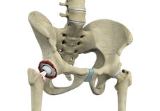
During total hip replacement, the damaged cartilage and bone are removed from the hip joint and replaced with artificial components. At times, hip replacement implants can wear out for various reasons and may need to be replaced with the help of a surgical procedure known as revision hip replacement surgery.

Reemplazo De Cadera Posterior Mínimamente Invasivo
El reemplazo total de cadera mínimamente invasivo es un procedimiento quirúrgico que se realiza a través de una o dos pequeñas incisiones en lugar de una sola incisión larga de 10 a 12 pulgadas, como en el enfoque tradicional.

Reemplazo de cadera anterior
La cirugía de reemplazo de cadera anterior directa es un procedimiento mínimamente invasivo en el que el cirujano de cadera reemplaza la articulación sin cortar ningún músculo o tendón.

Revisión de Cadera
Los implantes de una cirugía de reemplazo total de cadera pueden desgastarse por varias razones y necesitar ser reemplazados. El reemplazo de cadera de revisión es un procedimiento quirúrgico que sustituye total o parcialmente una articulación de cadera previamente implantada por una nueva articulación artificial de cadera.
